
Draw the structure of retina and explain the mechanism of vision in mammalian eye. (IAS 2022/15 Marks)
Draw the structure of retina and explain the mechanism of vision in mammalian eye. (IAS 2022/15 Marks)

Introduction
The retina is a complex structure located at the back of the eye that plays a crucial role in the process of vision in mammals. It contains specialized cells that are responsible for detecting light and converting it into electrical signals that can be interpreted by the brain.
Structure of the Retina
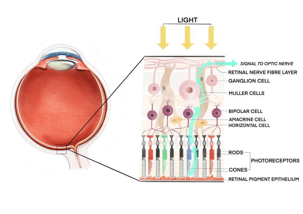
The retina is made up of several distinct layers that play specific roles in the process of vision:
- Outer Layer (Retinal Pigment Epithelium):
- Contains pigmented cells that absorb excess light and prevent scattering.
- Essential for the recycling of visual pigments.
- Photoreceptor Layer:
- Rod Cells: Responsible for vision in low light (scotopic vision). They do not detect color but are highly sensitive to light.
- Cone Cells: Responsible for color vision and detailed visual acuity. There are three types of cones sensitive to different wavelengths (red, green, and blue).
- Outer Plexiform Layer: Contains synapses between photoreceptor cells (rods and cones) and bipolar cells.
- Bipolar Cell Layer: Bipolar cells transmit signals from the photoreceptors to the ganglion cells.
- Ganglion Cell Layer: The axons of these cells form the optic nerve, which transmits visual information to the brain.
- Inner Plexiform Layer: Contains synapses between bipolar cells and ganglion cells.
- Optic Nerve Head (Blind Spot): The point where the optic nerve exits the eye. There are no photoreceptor cells here, resulting in a blind spot in the visual field.
Mechanism of Vision in Mammalian Eye
- Light Entry: Light enters the eye through the cornea, passes through the pupil (controlled by the iris), and is focused by the lens onto the retina.
- Light Absorption by Photoreceptors:
- When light strikes the retina, it is absorbed by photopigments in the rod and cone cells.
- In rods, the pigment rhodopsin absorbs light, while cones contain opsins that respond to different wavelengths of light corresponding to color perception.
- Phototransduction:
- The absorbed light causes a chemical change in the photopigment, triggering a cascade of reactions that result in the conversion of light into an electrical signal.
- In the case of rods, this leads to a change in membrane potential, known as hyperpolarization.
- Signal Transmission:
- The electrical signals generated in the photoreceptors are transmitted to bipolar cells, then to ganglion cells.
- Ganglion cell axons converge at the optic nerve, forming the visual pathway.
- Visual Information Processing: The visual signals travel via the optic nerve to the brain, specifically to the visual cortex of the occipital lobe, where they are processed and interpreted as visual images.
Conclusion
The structure of the retina and the mechanism of vision in the mammalian eye are intricate processes that involve the coordination of various specialized cells. By studying the structure and function of the retina, we can further our knowledge of vision and potentially develop new treatments for vision-related disorders.