
Draw a suitable diagram and explain the structure of human eye. (IAS 2022/15 Marks Marks)
Draw a suitable diagram and explain the structure of human eye. (IAS 2022/15 Marks Marks)

Introduction
The human eye is a complex organ responsible for the sense of sight. It is made up of various structures that work together to capture and process visual information.
Structure of the Human Eye:
- The human eye is a complex organ responsible for vision. It receives light and converts it into electrical signals, which are interpreted by the brain to form images.
- It is composed of various structures that work together to focus light, detect visual stimuli, and send signals to the brain.
- The study of the eye is essential in fields like zoology, biology, and medicine due to its importance in sensory functions.
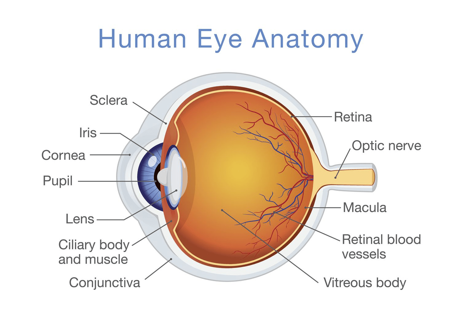
Key Parts of the Human Eye:
- Cornea:
- Transparent, dome-shaped outer layer.
- Acts as the eye's outermost lens and plays a significant role in focusing light.
- Provides protection to the internal structures.
- Anterior Chamber:
- Located between the cornea and the iris.
- Contains aqueous humor, a fluid that nourishes the eye and maintains intraocular pressure.
- Iris:
- The colored part of the eye.
- Controls the size of the pupil, regulating the amount of light that enters the eye.
- Contains muscles that adjust the pupil size in response to light conditions (dilates in dim light, contracts in bright light).
- Pupil:
- The black circular opening in the center of the iris.
- Controls the amount of light entering the eye.
- Lens:
- Transparent, flexible, and convex.
- Focuses light onto the retina, aiding in clear vision.
- Works in tandem with the cornea to focus objects at various distances (accommodation).
- Ciliary Body:
- Located behind the iris.
- Contains ciliary muscles that control the shape of the lens for focusing.
- Also produces aqueous humor to maintain the anterior chamber's pressure.
- Retina:
- The light-sensitive layer at the back of the eye.
- Contains photoreceptor cells (rods and cones) that detect light and color.
- Converts light into electrical signals that are transmitted to the brain via the optic nerve.
- Macula:
- A small area at the center of the retina.
- Responsible for central vision and fine detail.
- Contains a high concentration of cones (color-sensitive photoreceptors).
- Optic Nerve:
- Transmits visual information from the retina to the brain for processing.
- Forms the pathway for visual stimuli to be interpreted as images.
- Vitreous Humor:
- The clear, gel-like substance filling the space between the lens and retina.
- Helps maintain the eye's shape and allows light to pass through to the retina.
- Sclera:
- The white, outer layer of the eye.
- Provides structural support and protection.
- The sclera also serves as the attachment site for eye muscles.
Conclusion
The human eye is a remarkable organ that allows us to see and perceive the world around us. Its complex structure and functioning highlight the intricate design of living organisms, showcasing the wonders of zoology.